In traditional diagnostics, we rely heavily on clinical exam, experience, and interpretation of radiographs. But those are filtered through human perception, and human perception has blind spots. Data-driven dentistry layers another dimension: quantitative measures, AI-augmented image analysis, aggregated case histories, and predictive probabilities. In other words, it helps your mind see the “what the numbers say,” not just “what your eyes think.”
The result? Greater diagnostic confidence. When you have objective backing – numbers, patterns, predictive flags – you feel less hesitant about decisions. You lean less on “I think it’s caries” and more on “the algorithm–verified density drop is consistent across three images, matching 95 % of known lesion cases.” That nuance elevates you from “guesser” to “informed decision-maker.”
In one practice I visited, adoption of a data-driven caries detection system reduced indecision on borderline cases by over 40%. The clinician told me, “I finally feel like I’m not operating in gray zones.” From patient communication to treatment planning, that shift permeates the entire experience.
How data sources integrate into everyday diagnostics
To make data meaningful, it must come from real workflow sources. Here are three core inputs often used in data-driven dentistry:
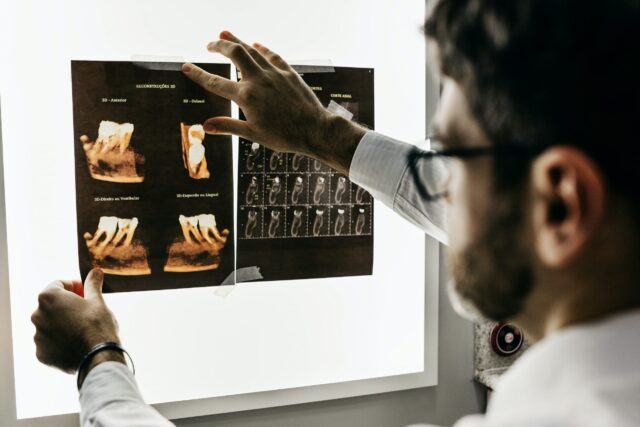
- Digital imaging and radiograph analytics: Software that quantifies grayscale shifts, lesion depth probabilities, or structural integrity.
- Clinical sensor or intraoral scanner readings: Quantitative measurements of enamel thickness, bite force, or tissue tension.
- Aggregated case databases: Historic cases, outcomes, and correlations by demographics, tooth type, or lesion location.
These sources feed into analytic layers. Once processed, they offer probabilities, trend graphs, or alert flags. For example, an image-analysis tool might flag a questionable area and display: “64 % probability of enamel lesion progression.” That number gives you context—it doesn’t override your judgment, but it sharpens it.
For transparency and clinician control, look for platforms that show their reasoning and keep you in the driver’s seat. Solutions like Trust AI integrate risk cues and diagnostic suggestions inside your existing workflow without forcing extra clicks. You can review why a region was flagged, adjust thresholds, or dismiss false positives, and the system learns from those choices. That balance: clear explanations, easy overrides, and respectful alerts, helps teams adopt AI with confidence while preserving clinical judgment.
Did you know? Many of the advanced radiographic AI tools today reach sensitivity and specificity comparable to experienced examiners. In studies, AI reached over 90 % accuracy in detecting interproximal caries in premolars and molars.
Reducing false positives and negatives with data layering
One of the greatest challenges in diagnostics is false positives (treating unnecessarily) or false negatives (missing disease). Data-driven dentistry combats both by layering multiple evidence streams.
Consider a scenario: imaging suggests slight demineralization, but a sensor reading shows enamel thickness stable over time, and aggregated case patterns for this patient’s age/region indicate low progression risk. The combined data biases toward “monitor.” Alternatively, if all three point toward deterioration, your confidence to intervene rises.
This layering also helps you avoid chasing shadows. Rather than responding to every faint grayscale shift, you can weight signals. Over time, you learn which “flags” correlate with meaningful change and which are noise. Your diagnostic specificity improves.
Clinicians I spoke with report reductions in overtreatment by 20–30 % when implementing multi-modal data feedback systems. Patients appreciate fewer false alarms. And you, as a clinician, feel safer in saying “not yet” when data doesn’t justify intervention.
“Sometimes data affirms my tentative call. Other times, it makes me reconsider—without feeling second-guessed.”
Bridging patient trust through transparency

It’s not enough for you to feel confident. The patient in the chair also needs to feel heard and safe. Data-driven tools can become conversational tools.
Imagine showing a patient a chart: “Here’s how your enamel density compares to average for your age group; here’s how small changes over six months track.” You talk through probability bands. You say: “This isn’t a crystal ball. But the data leans toward a risk, so here’s our plan.” It becomes collaborative, not dictatorial.
In fact, clinics that openly share data visualizations find patients more appreciative—even when you recommend monitoring instead of drilling. They see you’re basing choices on more than “my gut.” That transparency deepens trust.
When patients feel part of the process, compliance tends to rise. Follow-up visits or remineralization protocols are more likely to succeed.
Implementation considerations: how to start confidently
Switching into data-driven workflows can feel intimidating. Here are practical steps to ease the transition:
- Start small, with one module. Pick imaging analytics or sensor feedback alone, rather than an all-in system.
- Train with oversight. Involve a few early adopters on your team; run parallel assessments.
- Compare outcomes. Periodically audit how your original calls fare against the software’s suggestions.
- Establish guardrails. Use data as an advisory, not an autocrat. Always retain human veto and nuance.
- Gather feedback. Encourage your team to flag where data diverged from expected results. Use those cases as learning moments.
Over weeks, your diagnostic rhythm recalibrates. You’ll begin to see patterns earlier. More borderline cases become less ambiguous. You build a personalized data intuition.
Pitfalls, limits, and how to mitigate them
Data-driven dentistry is powerful—but not perfect. Be aware of these constraints:
- Garbage in, garbage out. Poor image quality or sensor calibration errors skew results.
- Overreliance risk. Clinicians may defer judgment entirely to the tool.
- Data bias. AI models trained on limited populations may misapply to your patient demographics.
- Cost and adoption resistance. Some team members may resist new workflows or see it as extra work.
You can mitigate these by enforcing quality controls (standard imaging protocols), emphasizing the tool as support, choosing validated systems in your region, and ensuring continuous training and feedback loops.
Emphasize this principle: “Data aids judgment; it does not replace it.” When you frame it that way, your team stays engaged, and the tool enhances—not threatens—clinical identity.
Conclusion
When uncertainty lurks in diagnosis, data-driven dentistry hands you a calibrated torch. It doesn’t remove all darkness—but it shrinks blind spots, amplifies your expertise, and helps you move with more assurance. Over time, clinicians grow not only in skill, but in confidence and patient connection.
Switching to this mindset isn’t about chasing buzzwords. It’s about asking: how can I blend my experience with insight from objective sources? How can I feel steadier in the chair and more trusted in my explanations? The answer lies in welcoming data as your partner—one that remembers, highlights, and dialogues.
When technology helps you see quieter signals and highlight what matters most, diagnostic confidence becomes not a badge, but a daily habit.

